Structure of protein from the visual cycle (RBP3) is solved
Reading time: about 7 minuts
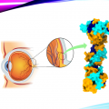
Proteins are fundamental biomolecules that perform a broad range of vital functions within the human body. They serve as an essential structural and functional component of cells, tissues and organs participating in processes ranging from basic cellular mechanisms such as DNA replication to complex physiological functions, including those involved in visual perception. In the visual system, proteins are important and crucial for light detection, the biosynthesis of photopigments in photoreceptor cells and intracellular signal transduction. Dysregulation or any kind of mutation in these proteins can disrupt the normal vision process and lead to various vision-related diseases. Recently, researchers from the Institute of Physical Chemistry Polish Academy of Sciences – International Centre for Translational Eye Research (ICTER) described the structural insight into the RBP3 protein improving our understanding of the visual cycle and its connection to retinal diseases.
Our natural optical detector – the eye is a fascinating and complex organ that helps us see the world around us. The proper functioning of this amazing organ requires the engagement of many different molecules. The vision starts with the retina which is a thin tissue layer covering the back of the eyeball. It works as the platform for many tiny cones and rods that are light-capturing photoactive detectors called photoreceptors. They convert the light into electrical signals that are sent to the brain through the neural system to interpret it as an image. Photoreceptors rely on the specific molecule 11-cis-retinal (11c-RAL), a light-absorbing molecule, which binds to opsin proteins (like rhodopsin) that initiate the visual process by converting light into electrical signals. When photons are absorbed, a cascade of chemical reactions, including the isomerization of 11-cis-retinal (11cRAL) to all-trans-retinal, initiates vision. To enable continued vision, the 11cRAL must be continuously regenerated through a process called the visual cycle. Here the story begins...
This is where another molecule enters the picture. That is Retinol-binding protein 3 (RBP3), a special protein located in the intercellular matrix that maintains the proper functioning of the visual cycle. RBP3 works as a transporter of retinoids between photoreceptors and retinal pigment epithelium cells and is also known to bind some important fatty acids. It shuttles crucial molecules back and forth from the photoreceptors making the visual pigments ready for the multiple reactions under the photons triggering.
The severity of diabetic retinopathy, an eye disease associated with diabetes, is associated with decreased levels of RBP3, and leads to progressive vision loss.
As RBP3 interacts with receptors like the glucose transporter 1 (GLUT1) and vascular endothelial growth factor (VEGF), been involved in blood vessel growth and cellular signaling in the eye. Disrupted RBP3 causes accumulation of retinal “waste products”, such as lipofuscin, which may cause oxidative damage to the RPE and photoreceptor cells. Besides diabetic retinopathy, RBP3 level disruption can also lead to retinitis pigmentosa, pan-retinal degeneration, and myopia.
Although the RBP3 connection with these diseases is well known, the mechanisms of the binding to retinoids to transport them are still not satisfactorily described. This mystery intrigued the international team of researchers led by dr. Humberto Fernandes from the Institute of Physical Chemistry, Polish Academy of Sciences – International Centre for Translational Eye Research (ICTER) to solve that mystery. They focused on the insight into the detailed structure of the RBP3 when it binds different retinoids and fatty acids. The main aim of their investigations was to overcome the lack of an experimental structural model for the native form of RBP3. To achieve this, the authors purified the porcine RBP3 (pRBP3) and analyzed its structure using cryo-electron microscopy (cryoEM), where data was collected under cryogenic conditions and after that data was refined by multiple steps and software to get the final 3D structure/model of the protein. Additionally, small-angle X-ray scattering (SAXS) was used to provide data on the conformation changes depending on the cargo molecules. Interestingly, the structure of the RBP3 can be elongated, or bent, suggesting the dynamic changes in the structure while docking its cargo.
“Based on previous knowledge of RBP3 properties and straightforward methods for isolation of the porcine variant of RBP3, we purified porcine RBP3, and obtained a protein with Förster resonance energy transfer behaviour analogous to other RBP3s. Through analysis of cryoEM data, we determined a structure at 3.67 Å resolution of the porcine RBP3 protein and observed conformational changes upon ligand binding.” – says dr. Humberto Fernandes
Experimental results enabled the determination of the 3D structure and revealed conformational changes upon binding to its ligand as a step forward in the insight into the RBP3 functional mechanisms during the visual cycle.
RBP3 as a large molecule consisting of four retinoid-binding modules has long lost its original catalytic functionality, and it evolved to be a cargo transporter interacting with varieties of molecules and delivering retinoids and fatty acids in the eye.
Research findings show the protein changes employing its shape during the binding of different molecules that relates to the effectiveness of the interaction with the other molecules in the cargo and signaling. As an effect, the conformational changes may play a significant role in the regulation of the light conversion into the visual signals.
Dr. Fernandes remarks “In all measured parameters, the porcine variant mimics the more completely characterized bovine variant. The capacity of RBP3 to load different retinoids and fatty acids, the ability of the latter to displace the former and the conformational changes dependent on ligand identity might be the basis for the loading and unloading of retinoids (and potentially DHA) to the intended cell types bordering the IPM intercellular matrix. Thus, RBP3 complexes merit further investigation.”
Understanding the proteins including genetic mutations that affect the protein's behaviour, like studied RBP3, is crucial to describe the mechanisms of the processes that appear in retinal diseases. Revealing the detailed structure of this bioactive molecule is a milestone in the studies on the interactions with different proteins. The presented findings bring the bright light into potentially more effective and faster diagnostics, where the RBP3 molecule would work as the early-stage retinal disease development biomarker. What is more, it can help in the regulation of the RBP3 activity to develop treatments for the disruption of the visual process.
The work was supported Foundation for Polish Science co-financed by the European Union under European Funds for Smart Economy (FENG.02.01-IP.05-T005/23), and (MAB/2019/12) project within the International Research Agendas programme of the Foundation for Polish Science co-financed by the European Union under the European Regional Development Fund. It was also supported by the National Institutes of Health R01EY009339. The authors also acknowledge support to the Department of Ophthalmology Gavin Herbert Eye Institute at the University of California, Irvine from an unrestricted Research to Prevent Blindness award, from NIH core grant P30EY034070, support by MEYS CR (LM2023042) and European Regional Development Fund-Project "Innovation of Czech Infrastructure for Integrative Structural Biology" (No. CZ.02.01.01/00/23_015/0008175) and iNEXT-Discovery, project number 871037, funded by the Horizon 2020 program of the European Commission, the PASIFIC postdoctoral fellowship programme (Agreement No PAN.BFB.S.BDN.315.022.2022; Project No. DPE/2023/00007), this project has received funding from the European Union’s Horizon 2020 research and innovation programme under the Marie Skłodowska-Curie grant agreement No 847639 and from the Ministry of Science and Higher Education, and cryoEM training through the Wellcome/MRC-funded cryoEM training program (218785/Z/19/Z).
CONTACTS:
Dr. Humberto Fernandes
Institute of Physical Chemistry of the Polish Academy of Sciences
International Centre for Translational Eye Research (ICTER)
email: hfernandes@ichf.edu.pl
SCIENTIFIC PAPERS:
“CryoEM structure and small-angle X-ray scattering analyses of porcine retinol-binding protein 3”
Vineeta Kaushik, Luca Gessa, Nelam Kumar, Matyáš Pinkas, Mariusz Czarnocki-Cieciura, Krzysztof Palczewski, Jiří Nováček, Humberto Fernandes
Open Biology, 2025
DOI: https://doi.org/10.1098/rsob.240180
- Author: Dr Magdalena Osial
- Contact: magdalena@osial.eu
- Photo source: Grzegorz Krzyżewski
- Date: 16.04.2025


