How do we use STOC-T to assess ocular microcirculation?
Reading time: about 7 minuts

Like a complex network of highways, the retinal microcirculation is a hidden system that powers the life of the eye – delivering oxygen, nourishing tissues, and allowing cells to function without disruption. New research conducted by ICTER (International Centre for Translational Eye Research) scientists will enable us to track every “movement” on these microscopic roads using the STOC-T (Spatio-Temporal Optical Coherence Tomography) technique. This offers the opportunity to understand the mechanisms of retinal function and discover how microcirculation disorders herald the onset of neurological and ophthalmological diseases.
Retinal microcirculation and hemodynamics provide valuable information on neurovascular diseases, as many diseases of the central nervous system (CNS) can manifest themselves through changes in the retina. Given that about 80% of external information is processed through visual perception, understanding the structure and function of the retina, vascular hemodynamics, and neurovascular coupling (NVC) is of paramount importance.
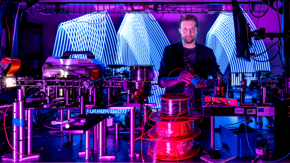
Now, ICTER scientists have used spatio-temporal optical coherence tomography (STOC-T) to assess retinal microcirculation. It turns out that the STOC-T technique, which uses fast near-infrared tomographic imaging, offers the possibility of visualizing even the smallest capillaries in real-time. Unlike other techniques, such as ocular angiography (angio-OCT) or Doppler tomography, STOC-T allows for obtaining 3D images of the entire structure of the retina and choroid with high temporal precision. Additionally, the use of digital aberration correction and a specially designed optical system allows for obtaining images unaffected by refractive errors, which is particularly important for imaging small structures, such as the mouse retina. The results were published in the journal Neurophotonics in a paper entitled "In vivo volumetric analysis of retinal vascular hemodynamics in mice with spatiotemporal optical coherence tomography."
What connects STOC-T and ocular microcirculation?
Spatio-temporal optical coherence tomography (STOC-T) is an advanced optical tomography method that allows for obtaining three-dimensional images of tissue microstructures in real time with high temporal resolution. In turn, ocular microcirculation is, broadly speaking, a network of small blood vessels supplying the retina and choroid, allowing for the proper functioning of photoreceptors. Adequate blood flow is essential for the delivery of oxygen and nutrients and the removal of metabolic products.
Retinal microcirculation research is becoming particularly important in the context of the increasing number of neurodegenerative and ophthalmological diseases. Disorders such as Alzheimer's disease, Parkinson's disease, and multiple sclerosis, as well as eye diseases such as glaucoma or diabetic retinopathy, are often associated with microcirculation disorders that can be visible at the retinal level even before neurological symptoms appear. The use of STOC-T allows precise monitoring of hemodynamic changes in the retina, which can help in the early detection of pathologies and the development of new therapeutic methods.Retinal blood flow can be quantitatively monitored in vivo using laser speckle flowgraphy (LSFG), a technique that generates angiographic contrast from speckle variance, enabling full-field (arbitrary units) blood flow measurements with high temporal resolution. Alternatively, laser Doppler flowmetry (LDF) can measure blood flow and mean velocity in relative units, and its extension, laser Doppler holography (LDH), can estimate pulsatile retinal flow in the lateral field of view (FOV) with millisecond resolution. These techniques cannot perform deep slices, making the influence of choroidal flow unclear. It would be beneficial to analyze choroidal hemodynamics separately from the internal retinal hemodynamics and with high temporal resolution, which is what STOC-T allows.
Groundbreaking observations and a chance for new therapeutic options
The study aimed to implement the STOC-T technique, previously developed by ICTER scientists, for monitoring retinal microcirculation and neurovascular coupling (NVC). Now, it was possible to obtain detailed images of different layers of the mouse retina, such as the neurofibrous layer (NFL), the inner plexiform layer (IPL), the inner and outer photoreceptor segments (IS/OS) and the choroid. These images allow for the observation of both larger blood vessels on the surface and the more complex network of capillaries in deeper layers, such as the IPL.
Analysis of the STOC-T signal amplitude allowed for the differentiation of arterial and venous pulsations in the mouse retina. In particular it was found that the pulsation in the venous vessels is delayed by an average of 29 milliseconds about the arteries, which allows for the identification of phase differences between these vessels. This pulsation time delay between arteries and veins is crucial for understanding the different roles these vessels play in microcirculation. STOC-T allows tracking of tissue displacements induced by the pulse wave as it travels through the retinal layers. These micromovements are measured in nanometers and observed mainly around arteries and veins, with modulation amplitudes ranging from 100 to 150 nanometers.
Measurement of the blood pulse wave velocity (0.35 mm/s) in the capillaries of the outer plexiform layer (OPL) and tissue displacements induced by vessel pulsation (up to 150 nm) provided data on the biomechanical properties of the different retinal layers. This analysis revealed differences in the biomechanical response to pulsation between layers, which is particularly valuable for NVC studies. Although the system is limited in recording pulse wave velocity in larger vessels due to the field of view and pulse wavelength, it remains highly effective in analyzing blood flow in capillaries.
Mapping of tissue shifts in time caused by vascular pulsation revealed that retinal layers exhibit periodic expansion and contraction synchronized with vascular pulsation. These observations, with an amplitude of 100-150 nm, provide important information on tissue elasticity and biomechanical properties of the retina, enabling further studies on neurodegenerative diseases in which the preservation of microcirculation and vascular elasticity may be impaired.
New quality in imaging of ocular hemodynamics
Studies conducted using the STOC-T technique provided detailed data on the hemodynamics and biomechanics of the mouse retina, which opens new diagnostic and therapeutic perspectives. The possibility of noninvasive monitoring of retinal blood flow and precise analysis of phase differences between venous and arterial pulsations may be crucial in the detection and treatment of many neurological and ophthalmological diseases. Ocular microcirculation, being a hidden highway supplying the retina with oxygen and nutrients, is crucial not only for the health of the eyes but also for the condition of the entire nervous system.
This publication is the result of the fruitful cooperation of three ICTER groups: POB, IDoc, and OBi, which emphasizes its interdisciplinary nature. These teams, combining their unique experiences and expertise, created the foundations for innovative solutions described in the publication. Such a combination of knowledge from different research areas is a key element in the search for innovative answers to contemporary challenges, which is one of the pillars of ICTER.
Authors of the paper "In vivo volumetric analysis of retinal vascular hemodynamics in mice with spatio-temporal optical coherence tomography": Piotr Węgrzyn, Wiktor Kulesza, Maciej Wielgo, Sławomir Tomczewski, Anna Galińska, Bartłomiej Bałamut, Katarzyna Kordecka, Onur Cetinkaya, Andrzej Foik, Robert J. Zawadzki, Dawid Borycki, Maciej Wojtkowski, Andrea Curatolo.
Author: Scientific Editor Marcin Powęska
Photos: Dr. Karol Karnowski
-----------------------------------------------------------------------------------------------------------------------------------
ICTER, or the International Centre for Translational Eye Research, is a research, development, and innovation centre at the Institute of Physical Chemistry, Polish Academy of Sciences (IChF), based in Warsaw, Poland. ICTER was established in 2019 to develop cutting-edge technologies to support the diagnosis and therapy of eye diseases, based on funding from the International Research Agendas Programme of the Foundation for Polish Science, co-financed by the European Union - European Regional Development Fund. The centre is currently implementing the IRAP FENG grant of the Foundation for Polish Science. In 2024, the IChF has won a prestigious grant under the Teaming for Excellence / WIDERA program of Horizon Europe, which will enable ICTER unit to develop into a European centre of excellence.
- Author: Marcin Powęska
- Photo source: Dr. Karol Karnowski
- Date: 18.12.2024


